选择你的语音


TurboScan自動化顯微影像快速掃瞄系統
TurboScan自動化顯微影像快速掃瞄系統
Solutions for Automated Microscopy and Imaging!!
![]()
產品應用簡介
Objective Imaging公司的自動化高速掃描玻片影像系統(Surveyor Automated Scanning and Imaging with Turboscan),
利用相機硬體觸發同步訊號方式,同時控制自動移動平台的移動速度,可在載台高速移動的狀態下進行連續影像擷取,實現顯微光學中高倍率、高解析、大範圍的影像拼圖掃瞄整合應用。
快速的拍照掃描整個玻片或樣品,讓樣品內的觀察目標,快速的呈現出完整樣貌,讓專業研究者可以在很短的時間內,
了解整個樣品細微結構與組織形態,解脫顯微鏡高倍圍觀察的視野侷限,適用病理組織切片、金相組織大範圍掃瞄和半導體晶圓大範圍掃瞄拼圖等應用需求。
TurboScan只需簡單的滑鼠點擊設定,便可以檢測任何想要觀察或確認的玻片位置,
對於研究者後續重覆檢測確認、研討、發佈等工作,都可以節省很多的時間。
利基於OASIS Automation Controller與特殊的相機觸發功能,TurboScan提供一套理想的自動化掃片解決方案,
包含樣品的定位、掃描、隨機拍照、自動化拍照存檔等等。
結合Prior SL160玻片自動載入機,可容納兩百片玻片,更可以達成大量的自動化玻片掃描工作,
適用於大量病理切片顯微影像掃描應用,對於有大量玻片顯微拍照的工作者,是最佳的選擇。
相關應用:玻片掃描、病理切片掃描
|
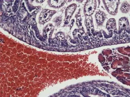
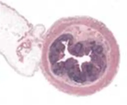
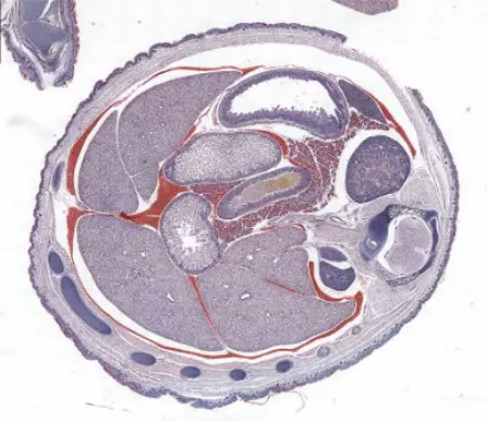
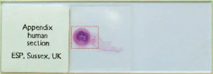
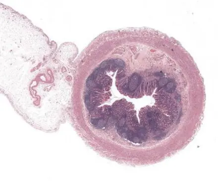
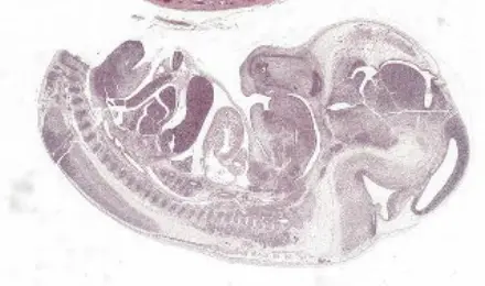
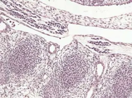
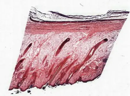
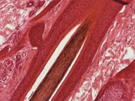
10X Objective / 1X C-mount Calibration: 0.46 µm/pixel Scan fields: 17 x 20, 340 total Size at full resolution: 340 x 4.2 MB = 1.4 GB Scan area: 10.6 x 9.2 mm = 98 sq. mm Scan time (snake scan with predictive focus and shading correction): 42 secs |
|
|
掃瞄後影像全覽
|
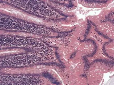
影像放大細節
|
|
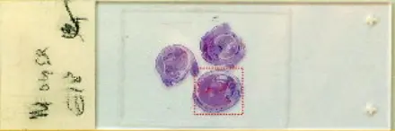
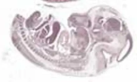
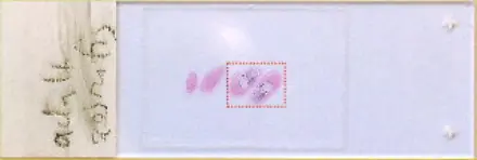
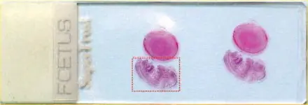
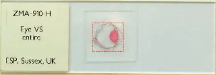
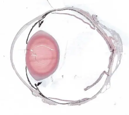
10X Objective / 1X C-mount Calibration: 0.46 µm/pixel Scan fields: 16 x 17, 272 total Size at full resolution: 272 x 4.2 MB = 1.2 GB Scan area: 9.2 x 7.3 mm = 67 sq. mm Scan time (snake scan with predictive focus and shading correction): 37 secs |
 |
|
掃瞄後影像全覽
|
影像放大細節
|
|
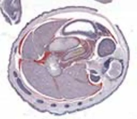
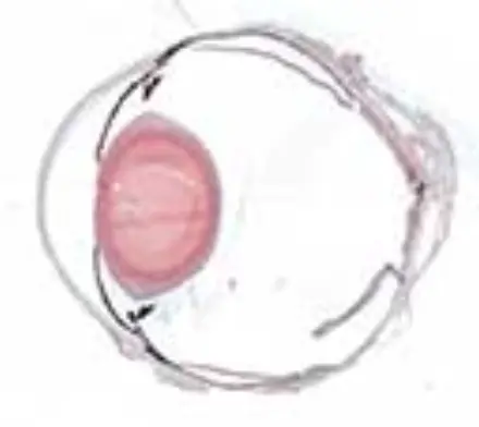
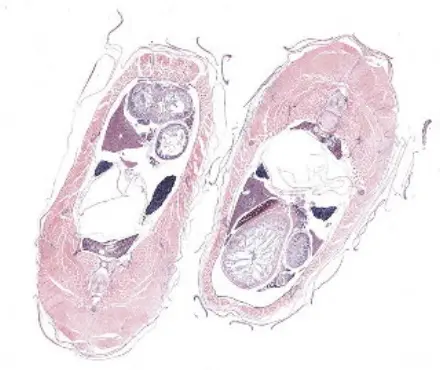
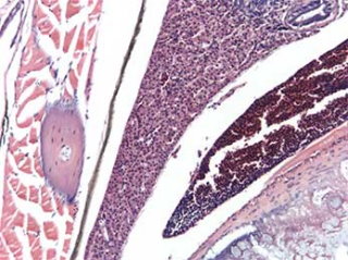
10X Objective / 1X C-mount Calibration: 0.46 µm/pixel Scan fields: 16 x 18, 288 total Size at full resolution: 288 x 4.2 MB = 1.25 GB Scan area: 10.1 x 8.2 mm = 83 sq. mm Scan time (raster scan with predictive focus and shading correction): 54 secs |
 |
|
掃瞄後影像全覽
|
影像放大細節
|
|
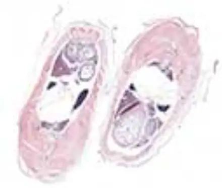
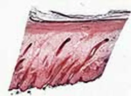
10X Objective / 1X C-mount Calibration: 0.46 µm/pixel Scan fields: 20 x 16, 320 total Size at full resolution: 320 x 4.2 MB = 1.3 GB Scan area: 12.5 x 7.4 mm = 93 sq. mm Scan time (snake scan with predictive focus and shading correction): 46 secs |
 |
|
掃瞄後影像全覽
|
影像放大細節
|
|
10X Objective / 1X C-mount Calibration: 0.46 µm/pixel Scan fields: 19 x 23, 437 total Size at full resolution: 437 x 4.2 MB = 1.8 GB Scan area: 11.9 x 10.6 mm = 126 sq. mm Scan time (raster scan with predictive focus and shading correction): 1 min 19 secs |
 |
|
掃瞄後影像全覽
|
影像放大細節
|

|
10X Objective / 1X C-mount Calibration: 0.46 µm/pixel Scan fields: 15 x 15, 225 total Size at full resolution: 225 x 4.2 MB = 0.95 GB Scan area: 9.4 x 6.9 mm = 65 sq. mm Scan time (raster scan with predictive focus and shading correction): 38 secs |
 |
|
掃瞄後影像全覽
|
影像放大細節
|
功能特點
Mosaic Acquisition
|
Specimen Map
|
Scan Patterns
|
Calibration
|
Camera Support
|
Microscope Automation
|
Export / Import
|
Automatic Focus |
如果您對自動化快速玻片影像掃瞄系統有興趣,歡迎來電洽詢,我們有專業的技術團隊可以提供您滿意的軟體客製化服務。
统一编号:28376870
服务邮箱: service@totalsmart.com.tw
地址:40744 台湾台中市西屯区河南路2段262号10楼之7
Monday – Friday
8:30 am – 5:30 pm CST
电话: +886-4-27080265 | 传真:+886-4-27080263
台北电话: +886-2-89924292 | 传真:+886-2-89929426
公司网站入口https://www.totalsmart.tw (繁體 ∣ 简体 ∣ English)
https://www.totalsmart.com.tw (繁體)
Copyright © 2026 Total-Smart Technology Co., Ltd. All Rights Reserved. | Privacy Policy | Terms of Use