选择你的语音


目鏡刻劃片 | 專業設計圖案
產品介紹
INTRODUCTION >> Microscope Eyepiece ReticlesSpray Droplet Sizing Reticle (Matthews) — NG30
For size and distribution assessments of aerosol droplets. Used in conjunction with a 40X Microscope for direct measurements of droplets from 50-400 micron diameters. The actual pattern sizes are 50, 100, 200, and 400 microns. W.H.O. (Details on Request) and G.A. Matthews. Imperial College.
Thompson — G23
For counting particles in any of three areas of known size. The graticule is calibrated in the same manner as a normal eyepiece scale. The result is then used to calculate the area of any square.
Chalkley Point Array — NG52
This is used to quickly determine the relationship of components to each other using random sampling. Curtis gives an example of its application, where a researcher may want to see whether or not a certain drug affects the volume proportion of cell types in a given organ. With this graticule the proportion of points lying over the image of one type of component is statistically proportional to the area occupied by that component. The 25 points of the array are placed over the field of view at random, so that a comparison can be made between the number of points touching one type of component, with the number touching the other type of component in each viewing. A series of observations will yield an increasingly accurate ratio of the comparative incidence of each type of particle.
Reference: A.S.C. Curtis. Medical and Biological Illustration, Vol. 10. pp 261-266. “Area and Volume Measurements by Random Sampling Methods.”
Pharmaceutical PSA Pattern — G57
This graticule was designed for the pharmaceutical industry. However, it is also useful where particle size considerations are restricted to 10mm and 25mm. Dots and circles give quick references for these two sizes. In addition, a scale is incorporated.
The microscope must be calibrated when ordering this graticule, such that the circle must equate to 1mm on the microscope stage.
Reference: The United State Pharmaceutical Conventions, Inc. Pharmaceutical Forum. Vol. 19 No 6.
Counting Pattern — NG14
Simple counting for geological and soil analysis.
Reference: L.G. Briarty. "Stereology: Methods for Quantitative Light and Electron Microscopy." Sci. Prog. Oxf. 1975 62; 1-32.
Lennox Grain Analysis — NG21
Reference: L.G. Briarty. "Stereology: Methods for Quantitative Light and Electron Microscopy." Sci. Prog. Oxf. 1975 62; 1-32.
Kotter Pattern — G48
For analysis of bitumous coal and anthracite.
Reference: I.S.O. 7404-4: 1988 (E). Methods for Analysis of Bitumous Coal and Anthracite. Part 4 and Methods of Determining Microlithotype Composition.
Normally used with 20x objective = calibration factor of 1. For use with 40x objective, specify calibration factor of 2. For 50x specify 2.5. For other objective magnifications, the reticle will need to be custom made.
Zeiss Integrating Eyepiece Disc 100 Points — G47
Similar to G49, but extended to 100 points, which are indexed.
Zeiss Integrating Disc 1 or Henning Reseau Pattern 25 Points — G49
Reference: Zeiss erkzeitschrft. No 30, page 80.
Integrating Eyepiece, G50
Stereology
In this simplest form, sterology is the science where information about a three dimensional object is obtained from only a two-dimensional section of that structure.
Measurements are usually made with these graticules in the following manner:
1. An adequate representation of sections of a specimen is obtained.
2. The graticule is superimposed upon the specimen (or micrograph/projected image of the section).
3. Finally, the interaction between the superimposed graticule and the test sections are recorded.
An overall introduction is given by: L.B. Brianrty. “Stereology: Methods for Quantitative Light and Electron Microscopy.”
The Mertz Graticule (36 Points) — NGM1
Used to estimate the three-dimensional surface areas or the surface density of a component in a given volume, when the component does not have a random orientation. It comprises a test system with parallel curved lines used for measuring the intersection of points.
Reference: W.A. Mertz. "Mikroskopic" Vol. 22 1967 pp 132-142.
Weibel 1 – NGW1
Consists of 15 lines of equal length connecting the verticals of a regular hexagonal point network.
Reference: E.R.Weibel Lab. Invest. Vol. 22 pp 131-152. "Principal and Methods for the Morphometric Study of the Lung and other Organs."
Weibel 2 – NGW2
Used when making a surface to volume ratio of a structure per mass unit. This graticule consists of a number of short lines with interruptions as long as the lines. Basically, the number of intersections falling over the short lines is counted and the number of endpoints falling on the end of the structure is determined.
Reference: E.R. Weibel, Journal of Microscopy Vol. 95. pp 373-378. Current Capabilities and Limitations of Available Sterrological Technique, point counting method.
Weibel 3 – GW3
Reference: E.R. Weibel, G.S. Kishtler & W.F. Scherle. 1986. J. Cell Biology. 30, 23.
包裝樣式
產品總覽
Products >> Microscope Eyepiece Reticles
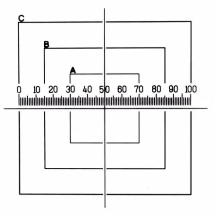
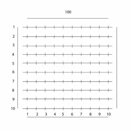
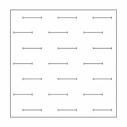
G23 Thompson for Dust Analysis
Eyepiece Reticle with Squares identified A, B and C and are sized 4mm, 7mm and 10mm. Horizontal Scale is 10mm subdivided into 100 divisions of 0.1mm (100µm).
For counting particles in any of three areas of known size. The graticle is calibrated in the same manner as a normal eyepiece scale. The result is then used to calculate the area of any square.
G48 Kotter Pattern
Eyepiece Reticle with Kotter pattern for analysis of bitumous coal and anthracite.
This reticle is normally used with a 20x objective and is sized accordingly. If other objective magnifications are used then we have to apply a calibration factor to ensure the pattern is the correct size. Please ask for a quotation for a custom made reticle. Reference: I.S.O. 7404-4: 1988 (E). Methods for Analysis of Bitumous Coal and Anthracite. Part 4 and Methods of Determining Microlithotype Composition.
G49 Henning Reseau Pattern - Zeiss Integrating Disc 1
Eyepiece Reticle with Henning Reseau Pattern (Zeiss Integrating Disc 1). 25 points on 6 horizontal lines.
G50 Integrating Eyepiece
Eyepiece Reticle with 100 point integrating pattern.
G57 Pharmaceutical PSA Pattern, IMA Reticle
Eyepiece Reticle for analysis in pharmaceutical applications
Often referred to as the Pharmaceutical PSA pattern, European Pharmacopeia pattern or IMA Reticle (USP 788).
Normally used on a microscope with 10x objective lens so that the main circle (10mm diameter) equates to 1mm at the stage. 10um and 25um dots and circles give quick reference to these sizes. A 10mm scale in 0.1mm divisions relates to 1mm in 0.01mm at the specimen stage when used with a 10x objective lens. If other objective magnifications are used, request a quote for a special calibration factor. We recommend that the PS8 Micrometer Scale with UKAS certificate of calibration is also purchased to calibrate the G57 reticle and provide measurement traceability for ISO.
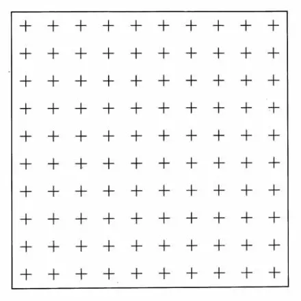
NG14 Counting Pattern
Eyepiece Reticle for Geological and Soil Analysis. 10mmx10mm square of 100 crosses in a 10x10 Array
Simple counting for geological and soil analysis. Reference: L.G.Briarty. “Stereology : Methods for Quantitative Light and Electron Microscopy.” Sci. Prog. Oxf. 1975 62; 1-32
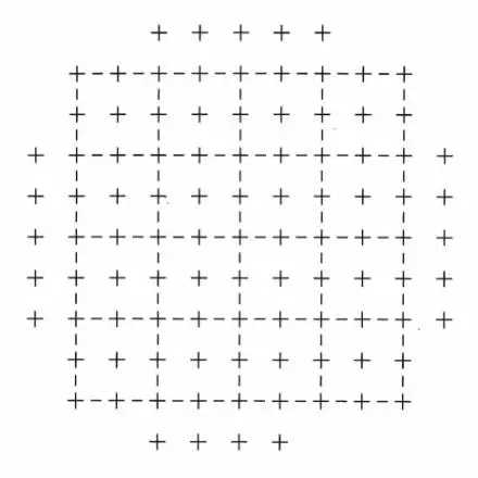
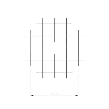
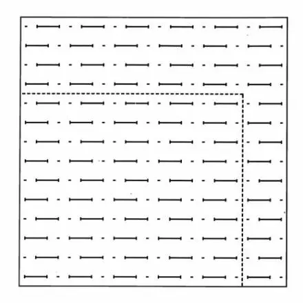
NG21 Lennox Grain Sizing
Eyepiece Reticle for Lennox for Grain Analysis. Array of dashed Squares and Crosses.
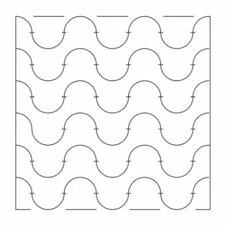
GW3 Weibel 3
Eyepiece Reticle with Weibel Type 3 pattern for Stereology. Pattern has a series of dots and horizontal dashes with alternate lines 'out of phase'.
Reference: E.R.Weibel, G.S.Kistler & W.F.Scherle. 1966. J.Cell Biology. 30,23.
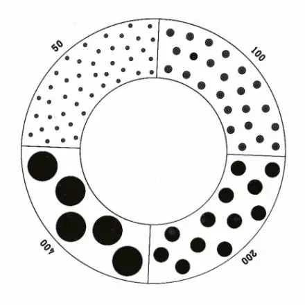
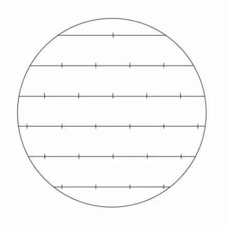
NG30 Matthews Spray Droplet Sizing
Eyepiece Reticle with Dots ranging from 0.050mm (50µm) to 0.400mm (400µm).
For size and distribution assessments of aerosol droplets. Used with 4x objective for direct measurements of droplets groups of 50, 100, 200 and 400 microns diameter. W.H.O. and G.A. Mathews. Imperial College.
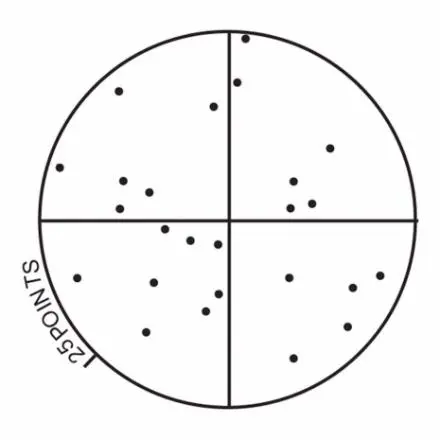
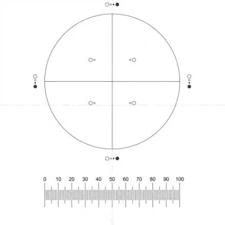
NG52 Chalkley Point Array
Eyepiece Reticle with Chalkley Point pattern. A single Circle divided into quarters and populated with 25 points (dots).
This is used to quickly determine the relationship of components to each other using random sampling. An example of its application is given by Curtis, where a researcher might want to see whether or not a certain drug affects the volume proportion of cell types in a given organ. With this reticle the proportion of points lying over the image of one type of component is statistically proportional to the area occupied by that component. The 25 points of the array are placed over the field of view at random, so that a comparison can be made between the number of points touching the one type of component, with the number touching the other type of component in each viewing. A series of observations will yield an increasingly accurate ratio of the comparative incidence of each type of particle. Ref. A.S.C.Curtis. Medical and Biological Illustration, Vol. 10. pp 261- 266. “Area and Volume Measurements by Random Sampling Methods”
NGM1 Mertz 36 Point
Eyepiece Reticle with Mertz pattern for Stereology
Used to estimate the three dimensional surface areas or the surface density of a component in a given volume, when the component does not have a random orientation. It comprises a test system with parallel curved lines used for measuring the intersection of points. Reference: W.A.Mertz. “ Mikroskopic” Vol. 22 1967 pp 132-142.
NGW1 Weibel 1
Eyepiece Reticle with Weibel Type 1 pattern for Stereology. 15 Lines of equal length connecting the certicals of a regular hexagon point network.
15 lines of equal length connecting the verticals of a regular hexagonal point network. Reference: E.R.Weibel Lab. Invest. Vol. 22 pp131-152. Principles and Methods for the Morphometric Study of the Lung and Other Organs.
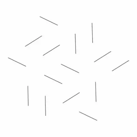
NGW2 Weibel 2
Eyepiece Reticle with Weibel Type 2 pattern for Stereology. Square area containing horizontal lines with spaces between of equal length.
Used when making a surface to volume ratio of a structure per mass unit. This reticle consists of a number of short lines with interruptions as long as the lines. Basically, the number of intersections falling over the short lines are counted and the number of endpoints falling on the end of the structure are determined. Reference: E.R.Weibel, Journal of Microscopy Vol. 95. Pp 373-378. Current Capabilities and Limitations of Available Stereological Techniques, point counting method.
统一编号:28376870
服务邮箱: service@totalsmart.com.tw
地址:40744 台湾台中市西屯区河南路2段262号10楼之7
Monday – Friday
8:30 am – 5:30 pm CST
电话: +886-4-27080265 | 传真:+886-4-27080263
台北电话: +886-2-89924292 | 传真:+886-2-89929426
公司网站入口https://www.totalsmart.tw (繁體 ∣ 简体 ∣ English)
https://www.totalsmart.com.tw (繁體)
Copyright © 2026 Total-Smart Technology Co., Ltd. All Rights Reserved. | Privacy Policy | Terms of Use